


Abdominal wall reconstruction
Overview
This term is used to describe techniques for repair of large complicated abdominal wall hernias.
Indications for its use include:
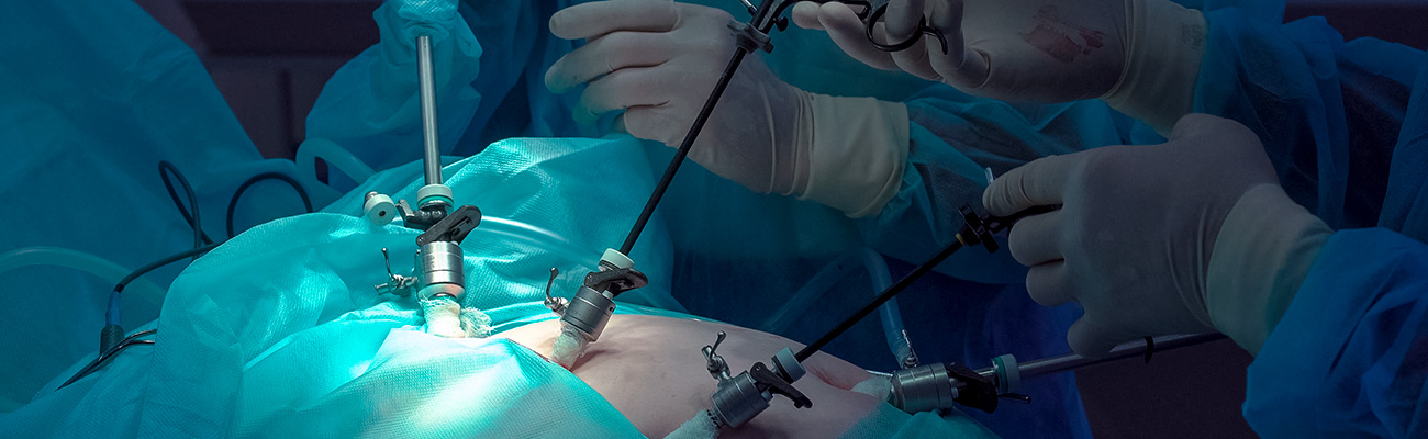
Abdominal wall reconstructions are typically done through an open technique, which involve a single incision / cut (normally up-and down and middle) on the abdominal wall. A layer of abdominal wall muscle is divided to allow increase mobility and ease of mobilisation of the hernia defect, to facilitate a tension free closure of the hernia defect.
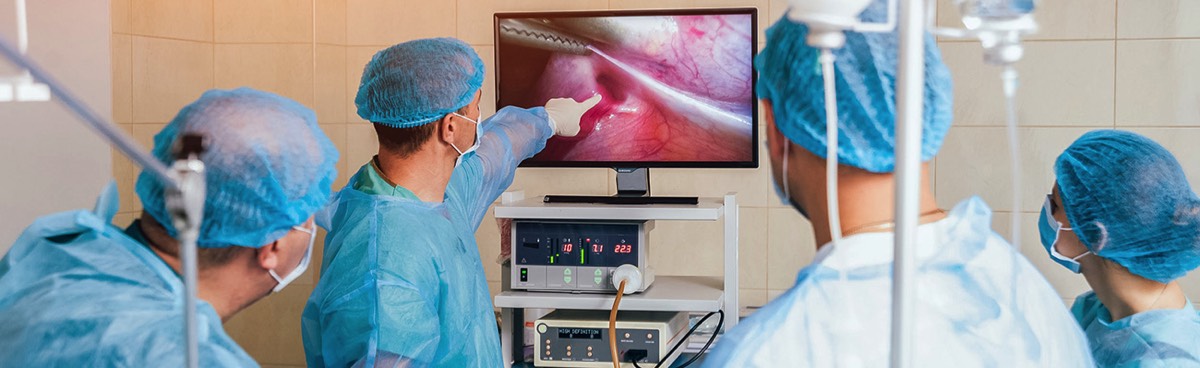
There are however increasing number of reported cases of abdominal wall reconstruction being done through the minimal invasive technique (‘keyhole’ surgery). This can be performed through laparoscopy or endoscopic approach.
There are two categories of abdominal wall reconstruction techniques:
Indications for its use include:
- Size of the hernia, which preclude the ability to approximate the edges of the hernia defect together without creating tension.
- Loss of muscular tissue
- Recurrent ventral hernias.
Abdominal wall reconstructions are typically done through an open technique, which involve a single incision / cut (normally up-and down and middle) on the abdominal wall. A layer of abdominal wall muscle is divided to allow increase mobility and ease of mobilisation of the hernia defect, to facilitate a tension free closure of the hernia defect.
There are however increasing number of reported cases of abdominal wall reconstruction being done through the minimal invasive technique (‘keyhole’ surgery). This can be performed through laparoscopy or endoscopic approach.
There are two categories of abdominal wall reconstruction techniques:
- Anterior component release – The external oblique muscle (front muscle on the side of the abdomen) is divided along the edge of the rectus abdominis muscle (‘six pack’ muscle)
- Post component release – The transversus abdominis muscle (back muscle on the side of the abdominal wall) is divided.

Procedure
With a hernia which has a large defect, the abdominal wall edges are widely separated. It is often difficult to bring the muscle edges together to close the hernia defect.
Even when it is possible to do so, the resultant repair will end up with a repair under high tension. This would result in a high rate of hernia recurrence.
Even when it is possible to do so, the resultant repair will end up with a repair under high tension. This would result in a high rate of hernia recurrence.

These hernias will require more advanced repair (abdominal wall reconstruction and abdominal wall component separation), involving incision on some layers of the abdominal wall muscle to allow further relaxation of the abdominal wall muscles to enable closure of the hernia defect.
These repairs may also be aided by utilising a Botox injection into the muscle a few weeks prior to the surgery.
These repairs may also be aided by utilising a Botox injection into the muscle a few weeks prior to the surgery.

Abdominal wall muscle.
Dr Ahmad’s experience
These component separation repairs are major complicated operations. They are typically only performed by a limited number of surgeons who have had dedicated training in this. Mr Ahmad has had considerable experience in this technique, with experience gained after spending time with Professor Todd Heniford of Carolinas Medical Centre, North Carolina—one of the premier hernia surgeons in the world.
He has also gained operative experience with Professor Miguel Garcia Urena in Madrid Spain.
He has also gained operative experience with Professor Miguel Garcia Urena in Madrid Spain.
Abdominal Wall Reconstruction/Component Separation Repair
These open procedures are used for repair of complicated ventral or incisional hernias.
An incision is made over the site of the hernia, and the hernia content is reduced into the abdominal cavity.
An incision is made over the site of the hernia, and the hernia content is reduced into the abdominal cavity.

Hernia reduced.
The deepest of the three muscles attached to the rectus abdominis is divided. This allows the hernia defect to be closed.

Muscle divided.
A large piece of mesh is then placed in the resultant space to cover and reinforce the hernia.

Mesh placement.
Postoperatively in the hospital there will be monitoring equipment used to monitor the patient’s progress.
Complications
- Wound infection.
- Haematoma/seroma—blood or fluid collection.
- Wound dehiscence (wound edges separating).
- Wound slow healing.
- Hernia recurrence.
- Other complications of major general surgery such as Deep Venous Thrombosis (DVT) and chest infection.
Complication rates can be calculated on individual patient’s basis based on the CeDAR application.
Postoperative care
These repairs are considered major surgery and will require you to stay in hospital for several days.
Please also see: Hernia – overview
Please also see: Hernia – overview
Clinical Associate Professor Hairul Ahmad
MB BS, FACEM (1999), FRACS, MS
Suite 12, Waikiki Specialist Centre,
217 Willmott Drive, Waikiki
Practice Details
Waikiki
Suite 12, Waikiki Specialist Centre,
217 Willmott Drive, Waikiki WA 6169
(08) 9592 2298
Fax: (08) 6314 1524
or email us
Office hours
9am–4pm Monday to Thursday
Affiliations


© 2024 All Rights Reserved. Content and images on this website are subject to copyright.
Email us | Sitemap | Disclaimer | Login