


Incisional hernia
See general information on hernia here.
This is a hernia that is associated with a previous abdominal incision. It can be open or laparoscopic (keyhole).
This is a hernia that is associated with a previous abdominal incision. It can be open or laparoscopic (keyhole).
Presentation
It can manifest itself at anytime after the surgery – even many years later. There may be:
- Lump/bulge – The lump is commonly reducible (easily pushed back into the abdomen). At this stage it is labelled a reducible hernia. It may progress to become an irreducible hernia (not able to be pushed back into abdomen).
- Pain/discomfort – The appearance of the lump is often associated with some discomfort or pain. This is often most noticeable with some physical activities. However, it may progress to cause pain even without physical activitiy.
- Strangulation – The hernia lump may rapidly increase in size and be associated with severe abdominal pain, protracted nausea and vomiting, bloating and distended abdomen. The lump may also be very tender when touched. These are features of a strangulated hernia. In this situation, the hernia content is being progressively being deprived of its blood supply. This is a surgical emergency and the patient is strongly advised to seek medical treatment without delay.

Incisional hernia.
Incisional hernia repair
The surgical approach to an incisional hernia repair is primarily dependent on the location and size of the hernia. The repair can be performed either by laparoscopic techniques or by the open method.
Repair of incisional hernia with small defect
Surgical repair: please see ventral hernia repair surgery.
Open procedure
An incision is made over the hernia.
The hernia sac and its content are dissected off the surrounding structures. The hernia is then reduced (pushed back into the abdomen). A mesh is placed under the muscle to cover the hernia defect. This mesh is typically anchored to the overlying muscle.
The defect is closed with sutures. A temporary drain may be needed. The skin is then sutured closed.
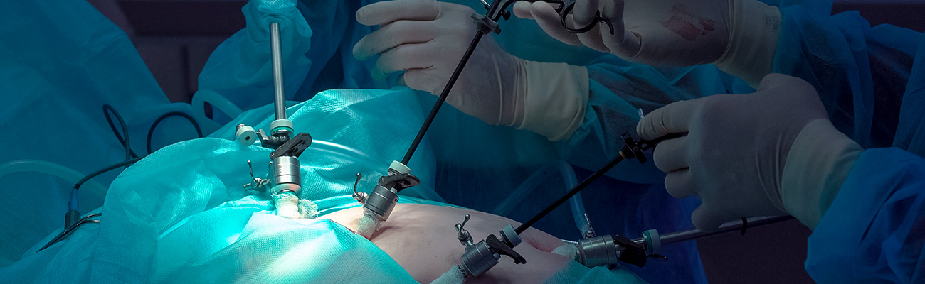
Laparoscopic procedure
Three incisions are made in the left abdominal wall, away from the hernia site. Gas is introduced into the abdominal cavity, elevating the abdominal wall away from the abdominal organs.
The hernia content is carefully dissected and reduced into the abdominal cavity leaving the hernia defect exposed.
The hernia defect is closed laparoscopically (from the inside). The hernia defect is then covered with mesh that is secured to overlying muscle with special sutures and clips. Skin is closed with dissolvable sutures.
Extended Totally ExtraPeritoneal Repair (eTEP)
This is a minimal invasive method of repairing the hernia by placing a mesh in the retromuscular space. The use of this space enables the placement of a large size mesh.

This is achieved through typically 3–4 small incisions. Because of this, there is quicker recovery from the surgery compared to the open technique (where the operation was performed through a large midline incision).

Repair of incisional hernia with large defect
Quite often in these hernias it is not possible to close the hernia defects without tension on the muscle repair. This will predispose to a high recurrence rate.
Therefore, for these hernias, the repairs are undertaken with the abdominal wall reconstruction approach.
See: Abdominal Wall Reconstruction/Abdominal Wall Component Separation
Therefore, for these hernias, the repairs are undertaken with the abdominal wall reconstruction approach.
See: Abdominal Wall Reconstruction/Abdominal Wall Component Separation
Possible complications
- Wound infection.
- Mesh infection.
- Wound haematoma/seroma.
- Hernia recurrence.
- Damage to abdominal organs (for laparoscopic repair).
Postoperative care
The patient should expect to have some discomfort for one to two weeks. This can be alleviated with some oral pain relief. A script will be given on discharge from hospital.
The patient is encouraged to be active. With restrictions of no heavy lifting for a minimum of 3 weeks. More detailed information will be discussed during your pre-operative consultation session.
Caution is advised when participating in exercise.
The patient is typically reviewed 2–3 weeks after the operation.
Please also see: Hernia – overview
Clinical Associate Professor Hairul Ahmad
MB BS, FACEM (1999), FRACS, MS
Suite 12, Waikiki Specialist Centre,
217 Willmott Drive, Waikiki
Practice Details
Waikiki
Suite 12, Waikiki Specialist Centre,
217 Willmott Drive, Waikiki WA 6169
(08) 9592 2298
Fax: (08) 6314 1524
or email us
Office hours
9am–4pm Monday to Thursday
Affiliations


© 2024 All Rights Reserved. Content and images on this website are subject to copyright.
Email us | Sitemap | Disclaimer | Login